The Doctor of Veterinary Medicine (DVM) program has now introduced a novel approach to assessment alignment in the histology module. This initiative is part of a broader shift towards a student-centred, active learning strategy, which aims to enhance student engagement and understanding through practical and interactive methods. By closely aligning assessments with learning activities, the program enhances the educational experience of our students. Histology is part of the Foundations of Veterinary Science A unit which has achieved most significant improvement in mean scores (>10% rise) in the Unit of Study Survey last semester.
Teaching sequence
The teaching sequence for each topic in the DVM1 histology module commences with allocated pre-work for the students to build a foundation before the lecture and practical activities. The pre-work comes in form of specified book-chapters for reading and short videos. The lecture then summarises the theory of a new topic, provides the framework and expected learning outcomes. Every lecture is followed by a guided microscopy session (1 hour) and practical activities (1 hour) to deepen students’ understanding and offer opportunities to have meaningful discussions with peers and academics. All assessments thereafter closely mirror the practical activities that students engage in during the semester.
In class activities and assessments
Drawing, annotations, and the early feedback task (EFT)
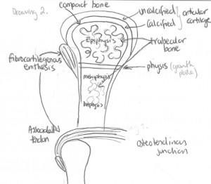
During the very first practical microscopy sessions in week 1 and 2, students practise drawing of tissue architecture with pen and paper to appreciate spatial arrangement of basic elements of tissues. This not only challenges their cognitive skills to recognise 3D structures on 2D images but also deepens their understanding of tissues architecture. The in-class EFT is a 40 minutes closed book exam at the end of week 3 and covers the introductory module and parts of the first musculoskeletal module. The EFT is worth 5% of the final mark and is similar in structure to the in-semester test (25%) and final exam (60%) which all include 10 minutes reading time.
In the EFT, in-semester test and final exam students are required to answer short answer questions and multiple choice questions by using pens, pencils, highlighters, but no electronic aids are permitted e.g. laptops, phones, smart watches. For the histology part of this early assessment students create a detailed and annotated drawing with pen and paper of a similar histological structure to that was previously analysed and discussed in class. In all further assessments throughout the semester students are allowed to draw tissue architecture to support their short and extended answers.
Essay writing and the ‘histo report’
In the microscopy practicals and associated activities in week 3 and 4, students practice describing normal tissue architecture first in simple, then in more sophisticated terminology through short essay writing. These activities are designed to prepare them to tackle both short and extended answer questions in the in-semester tests and final exam.
In addition, a new ‘histo report’, an individual assignment that counts for 15% of the final mark, has been introduced to allow students to show their acquired observational and analytical skills in their own pace and from home by providing online access to the microscopy platform Aiforia for 4 weeks until submission. In this assignment students analyse, describe, and interpret the tissue architecture of an organ of their choice but on a new and previously unknown tissue section.
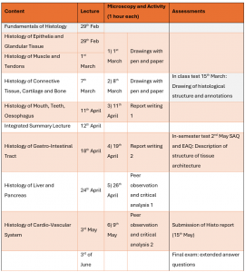
The histo report further comes with 2 alternatives, either a normal standard tissue approach or a modified tissue approach, in which students can unleash their creativity. In the modified tissue approach students are encouraged to modify their tissue section of choice to invent a more specialized form and function, which aligns with the concept of “tissue engineering.” This assignment pushes the boundaries of traditional histology and encourages innovative thinking. Close to 70 % of students chose the “modified tissue” option. Before the submission of the histo report, students were also able to present their tissue section to their peers in form of mini-presentations to receive immediate oral feedback.
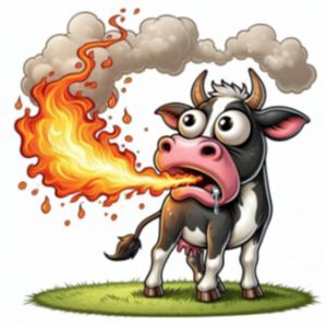
Examples of creative modified options were “Smokey Tauri” with methane sparking salivary glands, or “Anna the herbivore marsupial” with a bioluminescent nose pad that glows in the dark.

Student feedback
The creative assessments, such as the detailed drawings, histo reports, and especially the inventive “modified tissue” option, encouraged students to apply their knowledge in novel ways. This approach not only received highly positive feedback from students, who appreciated both the challenge and the creativity involved, but also improved Unit of Study Survey outcomes—as evidenced by the significant rise in mean scores.
- I absolutely loved the histology report and the modified option was super fun and original. It was the only assignment this semester that I actually enjoyed doing, across all of my classes.
- I had a fun time designing my cells 🙂 If I could do another “modified-cell” histo report in another class I would be looking forward to it.
- I loved the histology report and mini presentation and think that more of our histology grade should be in those forms.

