My experience with histology as a veterinary student was far from ideal. In a large lecture hall with 140 students, we had a single screen at the front displaying an image, a microscope with slides in front of us, and only three staff members to assist. To me, it all just looked like different shades of pink. Histology was a challenging subject for me, evidenced by the fact that I needed multiple attempts to pass it.
When I joined the Sydney School of Veterinary Science, I was tasked with taking over the histology teaching in the first year of the Doctor of Veterinary Medicine (DVM). Considering my own experience, this was a rather daunting thought at first, but I quickly realised that I could use my own experience to inform my teaching approach for this subject, and to hopefully provided a better learning experience to my students.
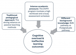
The DVM program enrols approximately 140 students annually, with about 40% being international students. The DVM is a postgraduate degree that requires, amongst other attributes, a strong academic record in undergraduate science courses for admission. The transition from undergraduate to the graduate degree is particularly challenging for students in the first year of the 4-year veterinary program (DVM1) where foundations are taught.
The heterogenous nature of the student cohort based on their entry pathway provides additional challenges.
There are two entry pathways to the DVM:
- Entry via an undergraduate pathway (80 students/year), with units specifically designed to provide vet-specific background knowledge e.g. Animal Structure and Function (AVBS2007). Students complete two years of undergraduate study at the University of Sydney and students with a weighted average mark (WAM) of 60 gain entry to DVM.
- Postgraduate entry (60 students/year). Student who completed a degree previously (Bachelor) and who meet pre-requisites (chemistry, biology, biochemistry).
Educational challenges

A gruelling schedule and a heavy reliance on passive teaching methods, such as lectures or lower cognitive tasks, result in an information-dense learning environment which can overwhelm students’ cognitive processing capacities, and lead to poor information retention, confusion, and a negative learning experience. This cognitive strain is often compounded by the lack of prior relevant veterinary knowledge due to the completion of a rather flexible and diverse undergraduate science degree and limited abilities in student’s self-directed learning.
Educational reforms
Threshold concepts
Introduced by Meyer and Land in 2006, this teaching approach has us identifying and defining fundamental concepts that form a barrier or threshold to allow for the deeper understanding of a subject. Threshold concepts are placed at the centre of all teaching, and students are provided with sufficient time to actively engage from different perspectives to build a solid foundation around those concepts.
For example, all organs are architecturally constructed by a set of specialised cells and their supportive stroma. Studying tissue “architecture” is a core concept of teaching histology in DVM. In the first few weeks of the histology module, students learn about functional elements of organs such as contractile, glandular, and epithelia tissues, and their structurally-supportive connective tissue. With those key elements in mind, students develop an understanding of their spatial arrangements and connections in organ systems through microscopy and through drawing.
Students have commented:
- I sincerely appreciated the thoughts put into the lectures, always trying to simplify very complex material so that it was as smooth as possible to take on new concepts.
- I discovered drawing to be one of the best tools for me to learn concepts.
Teaching for diversity
Inspired by Universal Design for Learning (UDL), the histology teaching team acknowledged different learning needs amongst the DVM student cohort. To transition from the existing purely didactic learning towards a blended-teaching approach, lectures were restructured not just to convey key concepts as a framework for learning, but also to incorporate various methods, such as live drawings to help students visualise concepts and apply material to real-world scenarios. A simple yet effective example involves using fruit to demonstrate how the appearance of an organ can vary significantly depending on how it is sectioned (cut).
The introduction of the cloud-based histology software (Aiforia) at the Sydney School of Veterinary Science allows histology sections used for teaching to be made available online. While students still attend in-person sessions to learn the physical handling and assessment of sections, Aiforia allows for the creation of short videos for each of the histology slides used in class. These videos, typically 5-10 minutes long, guide students through each histological section, consistently following the same systematic approach.
Students have said:
- I loved the free and open learning environment provided. I felt comfortable learning and developing my ways for effective learning.
- A fantastic job at structuring this course to engage students in the content. Understanding student needs, such as being visual learners, helps a great deal.
- There are so many ways provided to support our learnings (read, draw, write, listen etc). But they never forced us to adhere with certain ways or forced to do something. It was always a suggestion or part of activity.
Active learning
Supported by research, including Freeman et al. (2014), active learning strategies involve students more directly in their education through practical engagements, and higher cognitive task activities that encourage critical thinking and discussion, thereby fostering understanding and graduate qualities. The shift from a teacher-led to a student-centred approach helps in reducing cognitive overload by making learning more accessible and engaging.
For example, for each organ system students are provided with a 1 hour lecture only, but the emphasis lies on guided homework in form of bite-sized histology reading, videos, and interactive online tools that prepare students for the 1 hour of microscopy tutorial and 1 hour of student-led activities to explore tissue architecture in various ways, e.g. drawing, report writing, presentations, or peer discussions. The sequence of activities is designed to facilitate students’ growing cognitive skills throughout DVM1.
Students have noted:
- The histology portion of this course was one of my favourite units to date, and I think it is mainly because I felt as if I actually understood what I was learning and why. I enjoyed the structure of the course – Learning from lecture, pre-lab material, and then applying it once in the lab.
- I found the in class “quizzing” in the segments of the alimentary tract really helpful and engaging- if possible I would enjoy more quizzing throughout! This class has been extremely interesting and fun!
Transforming learning and engagement
These changes were designed to transform students’ learning experiences by promoting deeper understanding and engagement, thereby alleviating cognitive overload and enhancing mental well-being. The overwhelming positive feedback received from students, and their enthusiasm shown through engagement, made teaching histology extremely rewarding. This approach boosts their confidence and collaboration, enhancing their higher order cognitive skills. Additionally, I appreciated the endless opportunities to build trust and rapport with my students and develop a strong working relationship. I am confident that these simple educational reforms can be applied to other units.


