How often do your students memorise content but struggle to explain the deeper connections that make that knowledge meaningful? In veterinary education, understanding the intricate relationship between structure and function is a cornerstone of learning, yet these concepts are often taught in isolation.
For many of our students, often the missing link is understanding the role of “tissue architecture”—the organ-specific organisation of elements like epithelia, glands, connective tissue, and muscle that determines function. When students grasp how spatial arrangement creates functionality, and how disrupted architecture leads to dysfunction, they move beyond memorisation to genuine understanding.
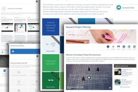
Our integrated veterinary histology-physiology course uses tissue architecture as the bridge between structure and function, enabling students to develop critical thinking skills and apply their knowledge to real veterinary scenarios. The approach also scaffolds learning through hands-on activities that incorporate visual, tactile, and collaborative elements.
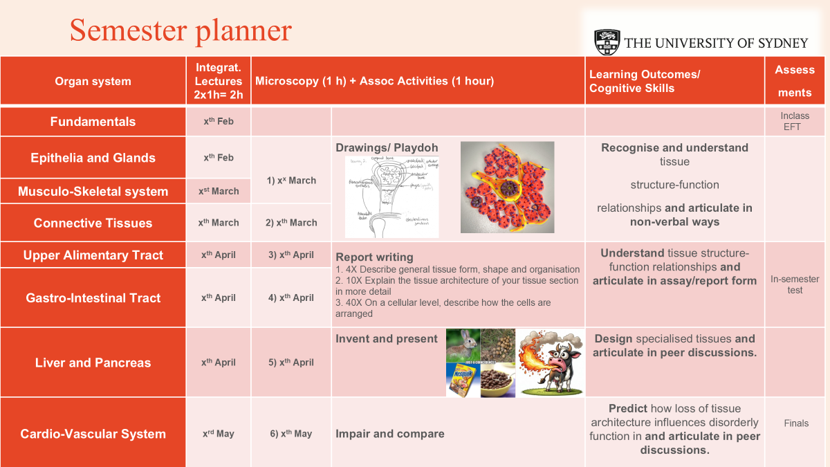
What we do: Scaffolding from observation to application
1) Drawing and annotating
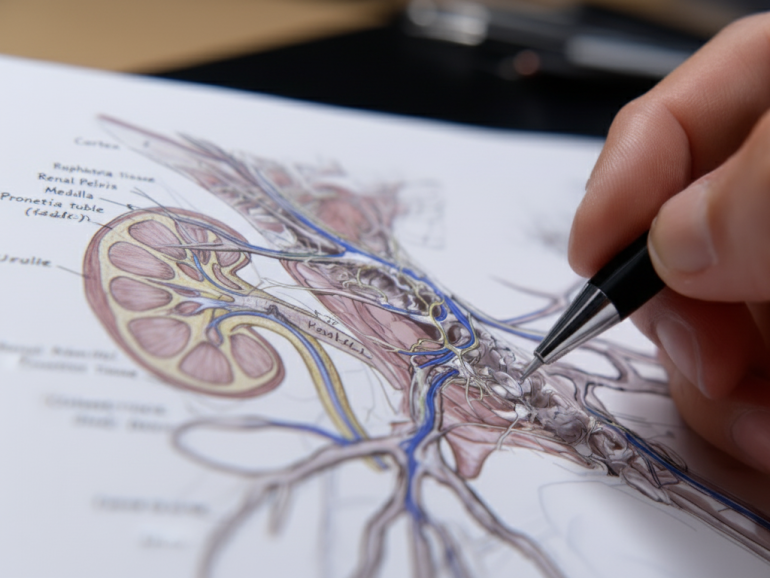
At the start of the course, students engage in drawing and annotating histological structures as a foundational activity. This helps them visually recognise tissue elements, understand spatial relationships of tissue architecture. These exercises promote observation skills and cognitive processing, making the transition to functional interpretation smoother. Most students choose to continue drawing throughout the semester.
2) Report writing
Building on their visual and spatial understanding, students then describe tissue architecture through short essays. This activity teaches them to articulate complex structures verbally, develops their ability to explain concepts clearly and concisely, preparing them for both assessments and professional report writing.
3) Tissue engineering with creativity
To deepen their understanding, students then participate in other hands-on activities where they design specialised tissues with “fantastic” functions using Play-Doh and present their ideas to peers. This exercise again reinforces spatial understanding, the application of structure-function concepts while developing communication, teamwork, and critical thinking skills. The tactile, creative approach (and getting to play with Play-Doh!) proves engaging and helps students think beyond traditional tissue functions.
3) Predicting functional implications
Finally, students are tasked with predicting the consequences of tissue derangements, linking structural changes to potential dysfunctions or disease. This develops critical reasoning skills and deepens understanding of tissue dynamics. Group discussions facilitate this capstone activity, helping students apply their theoretical knowledge to practical, clinical scenarios.
What we assess: Aligning assessment with in-class learning
Our assessments directly mirror the scaffolded activities, ensuring students are evaluated on the skills they’ve been developing:
- Histological section descriptions: Students describe tissue architecture at different magnifications.
- Functional interpretation: Students deduce tissue functionality based on structural arrangements.
- Predictive reasoning: Students justify how changes in tissue structures could lead to dysfunction or disease, integrating their knowledge of histology and physiology.
What do the students think?
Although this module is still in its early stages, feedback from students has been promising. Anonymous comments from unit surveys suggest that this approach improves student experience significantly, particularly through enhancing engagement and deep learning:
Caroline is used her remarkable teaching methods to make one of the hardest modules fun and easy to understand. I found this structure beneficial and it helped me retain information.
Student feedback, Unit of Study Survey responses, 2025
How can you adapt this approach?
Thinking about trying this approach? Here are my tips for getting started:
- Start small: Scaffold activities to build cognitive skills progressively – allow students time to develop confidence before moving to complex tasks. Diagrams and annotated drawings reduce complexity and support diverse learning needs.
- Incorporate hands-on elements and encourage creativity: Use tactile tools like Play-Doh and drawing materials to help students engage in multiple ways and give them choices. They will feel naturally drawn towards their favourite activity and engage with enthusiasm. Drawing and kneading clay has also been found as quite relaxing while actively learning.
- Emphasise communication: Include both verbal and written exercises to help students articulate complex concepts – particularly valuable for students developing academic English.
Final thoughts: Moving from memorisation to deep learning
This integrated approach demonstrates how breaking down barriers between related subjects/topics can transform student understanding. By using tissue architecture as a bridge concept, students moved from rote memorisation to meaningful learning that they can apply in clinical contexts.
The scaffolded activities – from drawing to predicting – show that when we align our teaching methods with how learning actually happens, students develop the critical thinking skills they need for professional practice. Most importantly, this approach is adaptable: the principle of using bridge concepts and hands-on scaffolding can work across disciplines where connecting structure to function matters.